At the office of Dr. Aaron Tropmann & Dr. Gary Oyster, we use advanced imaging tools to improve diagnostic confidence and patient outcomes. One of the cornerstones of our diagnostic suite is cone-beam computed tomography (CBCT), which provides three-dimensional views dentists rely on to plan treatments accurately and safely. CBCT complements clinical exams and conventional radiographs by revealing anatomic detail that can be critical to a successful result.
This page explains how CBCT works, why it matters in modern dental care, and the specific ways we integrate three-dimensional imaging into patient care. The goal is to give you clear, practical information so you can understand how CBCT supports precise diagnosis and treatment planning without wading into unnecessary technical jargon.
Cone-beam computed tomography uses a cone-shaped X-ray beam and a flat-panel detector to capture a series of images while the scanner rotates around the head. These images are reconstructed into a 3D volumetric dataset that can be viewed in multiple planes. The result is a compact, high-resolution model of teeth, jaws, sinuses, nerve canals, and adjacent structures.
Unlike traditional two-dimensional X-rays, CBCT removes many of the overlaps and distortions that can obscure important details. Clinicians can examine cross-sections or generate virtual slices at any angle, which makes it much easier to locate hidden pathology, evaluate bone volume, and assess spatial relationships before treatment. The flexibility of viewing options is especially helpful when standard radiographs are inconclusive.
Modern CBCT systems are designed specifically for dental use, with field-of-view options that limit exposure to the area of interest. That targeted approach reduces the amount of radiation relative to larger medical CT scans, while still delivering the level of detail dentists need for precise planning. The technology balances diagnostic value and patient safety, enabling better-informed decisions.
One of the most common uses for CBCT in dentistry is implant planning. Successful implant placement requires a thorough understanding of bone shape, density, and the location of vital structures such as the inferior alveolar nerve and the maxillary sinus. CBCT supplies the three-dimensional perspective that lets a clinician map implant positions with millimeter-level accuracy.
By visualizing the jaw in 3D, the treatment team can simulate implant placement virtually, select optimal implant sizes, and determine whether bone grafting or sinus modification is necessary. This preoperative insight reduces surprises during surgery and supports restorative-driven planning—placing implants where the final crown or bridge will function and look best.
CBCT also aids complex extractions, surgical exposure of impacted teeth, and orthognathic assessments. In each case, the detailed anatomy captured by the scan informs the sequence of care, helps anticipate potential complications, and supports a more efficient clinical workflow from diagnosis through follow-up.
CBCT is a versatile diagnostic tool that extends well beyond implant dentistry. Endodontists use 3D imaging to identify root fractures, accessory canals, and the full extent of periapical pathology that might not appear on 2D films. Oral surgeons rely on CBCT to evaluate cysts, pathology, and the relationship of lesions to adjacent structures.
Orthodontic and airway assessments also benefit from volumetric imaging. CBCT can reveal airway volume and narrowings that may contribute to sleep-disordered breathing evaluations. Orthodontists use three-dimensional data to evaluate tooth position, jaw asymmetries, and growth patterns when planning comprehensive movement or surgical-orthodontic cases.
Periodontists and restorative dentists use CBCT to measure bone levels around teeth, evaluate furcation involvements, and plan ridge preservation or augmentation. In every specialty, having a complete three-dimensional map of the anatomic area improves diagnostic accuracy and makes treatment recommendations more predictable.
Patient safety is a primary concern whenever ionizing radiation is involved. Dental CBCT units are engineered to minimize exposure by limiting the scanned volume and using optimized protocols for different clinical needs. When a CBCT scan is recommended, the expected diagnostic benefit is weighed against the radiation dose to ensure it is justified.
Our practice follows contemporary guidelines for imaging selection and dose reduction. We choose the smallest field of view that still answers the clinical question and adjust settings for each patient’s size and the area being evaluated. When conventional radiographs provide sufficient information, we use those instead of CBCT.
From a patient experience standpoint, CBCT scans are quick, noninvasive, and typically completed in under a minute of rotation time. The open design of dental CBCT machines is more comfortable than enclosed medical CT scanners for many patients, and there is no need for injections or intravenous contrast for routine dental imaging.
Generating a 3D scan is only the first step; accurate interpretation is essential to translate images into safe, effective treatment. Our clinicians review CBCT datasets using specialized software that enables measurements, simulated implant placement, and assessment of anatomy from multiple perspectives. This process transforms raw data into actionable clinical information.
When findings suggest issues outside the typical dental scope—such as suspicious soft-tissue lesions or signs that warrant medical evaluation—we coordinate care with appropriate specialists. CBCT helps us recognize when collaboration or referral is in the patient’s best interest, ensuring comprehensive management rather than piecemeal treatment.
We also use CBCT results to enhance patient communication. Visualizing anatomy in 3D helps patients understand the rationale for recommended procedures, potential risks, and expected outcomes. Clear visual explanation supports informed decision-making and a collaborative approach to care.
Incorporating CBCT into our diagnostic toolkit allows us to deliver treatment plans that are precise, predictable, and tailored to each patient's anatomy. If you have questions about whether CBCT is appropriate for your situation or how it would be used in your care, please contact us for more information. We are happy to discuss how three-dimensional imaging may help support your treatment goals.


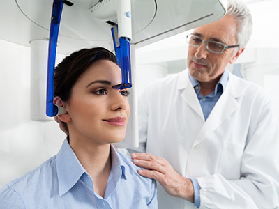
Cone-beam computed tomography, commonly called CBCT, is a dental imaging technique that captures volumetric, three-dimensional views of the teeth, jaws, sinuses and surrounding structures. The scanner rotates around the head and reconstructs a series of images into a single 3D dataset that can be viewed in multiple planes. This approach reveals spatial relationships and anatomic detail that are not visible on conventional two-dimensional radiographs.
CBCT removes many of the overlaps and distortions inherent to 2D films and allows clinicians to examine cross-sections or virtual slices at any angle for more precise diagnosis. At the office of Dr. Aaron Tropmann & Dr. Gary Oyster, CBCT is used to complement the clinical exam and traditional radiographs when additional anatomic clarity is needed. By combining image detail with clinical findings, the team can make more informed and predictable treatment decisions.
Traditional dental x-rays provide flat, two-dimensional images that are excellent for many routine diagnostic needs but can obscure depth and overlap of structures. CBCT produces a volumetric image that removes many of those overlaps, enabling clinicians to measure bone dimensions, locate nerve canals, and evaluate the position of roots or impacted teeth in three dimensions. This added spatial information is particularly valuable when 2D films are inconclusive or when planning surgery.
Unlike larger medical CT scanners, dental CBCT systems are designed with selectable fields of view so the imaging volume can be restricted to the area of interest, which improves efficiency and reduces exposure. The result is a more targeted diagnostic tool that balances image detail with patient safety. For routine care where a panoramic or periapical film answers the clinical question, those conventional images will still be used.
CBCT is a standard tool for modern implant planning because it shows bone height, width and the relationship to vital structures such as the inferior alveolar nerve and maxillary sinus. With a 3D dataset clinicians can virtually plan implant positions, evaluate bone volume, and select appropriate implant length and diameter with millimeter-level accuracy. This preoperative visualization reduces intraoperative surprises and helps tailor the procedure to each patient’s anatomy.
The volumetric data also allows for restorative-driven planning, meaning implants are positioned to support the final crown or bridge for optimal function and esthetics. When necessary, the scan helps determine whether bone grafting or sinus augmentation will be required before implant placement. Many practices also use CBCT datasets to fabricate surgical guides that further improve placement accuracy during surgery.
Your dentist may recommend CBCT when a clinical question requires three-dimensional information that cannot be resolved with conventional x-rays. Common indications include implant planning, evaluation of impacted or atypical tooth positions, assessment of complex root anatomy for endodontic cases, investigation of suspected cysts or pathology, and airway or orthognathic assessments. The recommendation is based on whether the additional anatomic detail will materially affect diagnosis or treatment planning.
CBCT is not used for routine preventive exams and is reserved for situations where the expected diagnostic benefit justifies the exposure. When a conventional radiograph provides the information needed, clinicians will choose the lower-exposure option instead. The decision to scan follows accepted imaging selection principles and is tailored to each patient and clinical scenario.
CBCT units designed for dental use deliver lower radiation doses than most medical CT scanners because they allow for smaller, targeted fields of view and optimized imaging protocols. Safety is managed by selecting the smallest field of view and appropriate exposure settings for the clinical task and the patient’s size. These choices help limit radiation while still providing diagnostically useful images.
Our practice follows the principle of ALARA — as low as reasonably achievable — and recommends CBCT only when the expected diagnostic benefit outweighs the exposure. When conventional radiographs can answer the clinical question, we use those alternatives. Patients with specific concerns about radiation are encouraged to discuss them so the team can explain the justification and safeguards used for their care.
A CBCT scan is a quick, noninvasive procedure that typically involves sitting or standing while the scanner rotates around your head to collect images. There are no injections or contrast agents for routine dental CBCT, and the machine’s open design tends to be more comfortable than enclosed medical CT units. The staff will position you carefully and ask you to remain still for the brief acquisition to avoid motion artifacts.
Most dental CBCT rotations are completed in under a minute, though total appointment time includes positioning and a short review with the clinician. You may be asked to remove jewelry or metal accessories from the head and neck area to minimize image artifacts. After the scan, the dataset is reviewed using specialized software to create the views needed for diagnosis and planning.
CBCT datasets are reviewed by the treating dentist or specialist using advanced imaging software that enables measurements, cross-sectional views and virtual treatment simulations. Many general dentists and specialists complete training in CBCT interpretation, and complex or unclear findings may be reviewed by a board-certified oral and maxillofacial radiologist. Accurate interpretation is essential to translate the images into safe, effective treatment recommendations.
Findings from the scan are integrated into a patient-specific plan that may include surgical guides, coordinated referrals, or modified operative approaches based on nearby anatomy. If the scan reveals concerns outside the dental scope, the team will recommend appropriate medical evaluation or specialist collaboration. Clear visualization also helps clinicians communicate treatment options and anticipated outcomes to patients.
Yes, CBCT can be an important adjunct in endodontics by revealing root fractures, extra or accessory canals, and the full extent of periapical pathology that might be hidden on 2D films. The 3D perspective enables clinicians to see complex root anatomy and canal curvature more clearly, which can influence decisions about retreatment, surgical intervention, or nonsurgical therapy. In certain persistent or atypical cases, CBCT changes the diagnostic picture and suggests a different treatment path.
That said, CBCT is used selectively for endodontic cases when the additional information will affect clinical management. Routine, straightforward root canal treatments are often completed with conventional radiographs. The treating dentist or endodontist will evaluate whether a scan is justified based on symptoms, prior imaging and clinical findings.
CBCT provides excellent bone and dental hard-tissue detail but has limitations in soft-tissue contrast compared with medical CT or MRI, so it is not a replacement for all medical imaging. Metal restorations can cause artifacts that degrade image quality in the area of interest, and small fields of view may miss pathology outside the scanned volume. Additionally, incidental findings can appear that require further evaluation or referral, which may add steps to care.
Because interpretation requires training, scans should be reviewed by clinicians familiar with CBCT artifacts and anatomic variants to avoid misdiagnosis. Careful case selection and clear communication about what the scan can and cannot show help manage expectations. When appropriate, the team will coordinate with specialists to ensure comprehensive assessment and follow-up.
Three-dimensional imaging makes anatomy tangible for both clinicians and patients, improving understanding of diagnoses, planned procedures and potential risks. Visualizing the jaw, tooth roots and adjacent structures in 3D supports restorative-driven planning and more predictable surgical execution, which can reduce intraoperative surprises and streamline care. Clear images also facilitate multidisciplinary coordination when multiple specialists are involved.
At the office of Dr. Aaron Tropmann & Dr. Gary Oyster, CBCT is used to enhance informed decision-making and help patients see the anatomic basis for recommended treatments. By pairing high-quality imaging with clinical expertise, the practice aims to deliver precise, individualized care that aligns with each patient's goals and anatomy. If you have questions about whether CBCT is appropriate for your situation, contact the office to discuss how 3D imaging might be used in your care.

Ready to book your next appointment or have a question for our team? We're here to help.
Connecting with our team is simple! Our friendly staff is here to help with appointment scheduling, answer any questions about your treatment options, and address any concerns you may have. Whether you prefer to give us a call, send an email, or fill out our convenient online contact form, we’re ready to assist you. Take the first step toward a healthier, brighter smile – reach out to us today and experience the difference compassionate, personalized dental care can make.

We serve the Raleigh-Durham area, including Cary, Apex, Chapel Hill, Morrisville, Wake Forest, North Hills, North Raleigh and more.
Back to top
