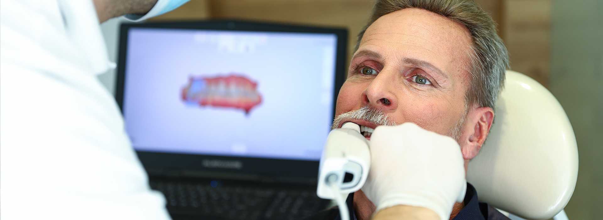
Digital impressions replace traditional putty-based molds with an intraoral scanner that captures high-resolution, three-dimensional images of teeth and soft tissues. During a scan, a wand-like device records a series of images that software stitches together to create an accurate digital model. That model can be viewed, rotated, and magnified on a monitor, giving both clinician and patient a clear, immediate view of the anatomy being treated.
Instead of producing a physical cast in the operatory, the scanner creates files (commonly in STL or other CAD-compatible formats) that are ready for design and fabrication. These digital files can be imported into dental design software for crowns, bridges, implant restorations, inlays/onlays, and orthodontic appliances. Because the data is inherently digital, it integrates smoothly with CAD/CAM workflows and modern dental laboratories.
The process minimizes the need for re-taking impressions: the clinician can review the scan in real time and address missing detail or soft-tissue interference right away. That immediate feedback helps ensure the captured anatomy is comprehensive and usable for the next steps in restoration or appliance fabrication.
One of the most noticeable benefits of digital impressions is patient comfort. Traditional impressions often require trays and viscous materials that can cause gagging or leave an unpleasant taste. Scanning is minimally invasive: the wand is moved around the mouth for short intervals while the patient breathes naturally and can communicate with the clinician throughout the capture.
Digital scanning also shortens chair time in many cases. Because scans can be reviewed immediately, adjustments are made on the spot rather than having to wait for a lab to identify an issue from a physical impression. For patients who need repeat impressions—such as for implant or prosthetic follow-ups—digital files can be recalled instead of repeating an uncomfortable impression procedure.
For patients with strong gag reflexes, medical sensitivities, or anxiety around traditional materials, intraoral scanning can make diagnostic and restorative appointments smoother and more tolerable without reducing clinical precision.
Digital impressions offer a level of dimensional stability that physical materials struggle to maintain. Impression materials can distort with time, temperature changes, or during shipping; digital scanners capture a direct optical map of the mouth that does not change once saved. This stability translates into restorations that fit more predictably and require fewer adjustments at delivery.
When restorations fit well, clinical outcomes improve: less chairside adjustment, stronger margins, and improved occlusal harmony. That precision benefits single crowns as well as multi-unit bridges and implant-supported prostheses, where small inaccuracies can compound across multiple units. Clinicians rely on digital scans to minimize these cumulative errors and to build restorations that function comfortably and esthetically.
High-resolution digital data also helps with communication between the dentist, dental laboratory, and specialist providers. Annotated scans and shared 3D models reduce ambiguity, so technicians can design and fabricate components that align closely with the clinician’s intent.
One practical advantage of digital impressions is the way they accelerate collaboration with dental laboratories. Digital files are transmitted electronically, eliminating the wait associated with shipping physical impressions and stone models. That direct transfer shortens turnaround times for lab-fabricated restorations while improving traceability and case documentation.
Many practices combine intraoral scanning with in-office milling or 3D printing to produce restorations the same day. For patients, this can mean fewer appointments and a quicker path from diagnosis to final restoration. In-office CAD/CAM systems use the same digital dataset as external labs, allowing clinicians to control design parameters and deliver a finished crown or provisional in a single visit when clinically appropriate.
Even when restorations are completed by an external lab, the initial speed and clarity of digital transmission reduce back-and-forth and help avoid costly remakes caused by ambiguous or distorted impressions.
Digital impressions become part of the patient’s permanent electronic record. These archives make it easy to compare changes over time, monitor wear, or plan staged treatments without re-imaging. Stored scans are a reliable baseline for future work—whether for restorative revisions, orthodontic movement, or implant planning.
In treatment planning, digital models work hand-in-hand with other technologies such as CBCT imaging and digital occlusal analysis to create coordinated, multidisciplinary plans. When implant restoration or complex prosthetic work is needed, the fusion of 3D scan data with radiographic imaging supports precise surgical guides and restorative-driven implant placement.
From an infection-control perspective, digital capture reduces the number of physical items that need sterilization or disposal. There is no impression tray to disinfect or impression material to manage, streamlining clinical protocols and lowering the handling of potentially contaminated materials while maintaining the highest standards of safety.
At the office of Dr. Aaron Tropmann & Dr. Gary Oyster, intraoral scanning is integrated into a digital-first approach to restorative and prosthetic dentistry. By embracing digital impressions, our clinicians improve predictability, reduce patient discomfort, and collaborate more efficiently with trusted laboratories and specialists.
To learn more about how digital impressions can benefit your next dental procedure, please contact us for more information.

Digital impressions use an intraoral scanner to capture high-resolution, three-dimensional images of teeth and soft tissues instead of traditional putty-based materials. A small wand records a sequence of images that software stitches into a precise digital model that can be rotated, magnified, and inspected on a monitor. These models are commonly saved in CAD-compatible formats such as STL and are ready for design and fabrication by the clinician or laboratory.
The digital workflow lets clinicians review scans in real time and correct any missing detail immediately, reducing the need for repeat captures. Because the data is inherently digital, it integrates directly with CAD/CAM systems, digital labs, and milling or 3D-printing equipment. This immediate feedback and compatibility help streamline restorative and appliance workflows from diagnosis to delivery.
Digital scanning eliminates the need for bulky impression trays and viscous materials that often provoke gagging, discomfort, or unpleasant tastes for patients. The process is minimally invasive: a wand is gently moved around the mouth while the patient breathes normally and communicates with the clinician throughout the capture. Short capture times and immediate review of the scan further reduce chairside anxiety and exposure to unfamiliar materials.
For patients with a strong gag reflex, medical sensitivities, or dental anxiety, intraoral scanning can make diagnostic and restorative appointments more tolerable without sacrificing accuracy. Scans can also be recalled later for follow-up work, avoiding repeated impressions and additional discomfort. That convenience often translates into a smoother and more positive clinical experience for many patients.
Digital impressions capture a direct optical map of the mouth that does not change once saved, whereas physical impression materials can distort over time, with temperature changes, or during shipping. The dimensional stability of a digital file reduces cumulative errors, which is particularly important for single crowns, multi-unit bridges, and implant-supported restorations where small inaccuracies can compound. High-resolution scan data supports precise margins, occlusion, and contact relationships that reduce chairside adjustments.
Because clinicians can review and annotate scans, communication with dental laboratories and specialists becomes clearer and less ambiguous. This improved coordination lowers the chance of remakes and ensures restorations align closely with the clinician’s plan. Overall, predictable fit and reduced adjustments lead to better long-term clinical outcomes.
Yes. Digital files are transmitted electronically to dental laboratories, eliminating shipping delays associated with physical impressions and stone models. Electronic transfer shortens turnaround times and improves traceability and case documentation, allowing labs to begin design and fabrication sooner. When combined with in-office milling or 3D printing, the same digital dataset can be used to produce provisional or definitive restorations in a single visit when clinically appropriate.
Even when external labs complete the final restoration, the clarity and speed of digital transmission reduce back-and-forth communication and the risk of remakes due to distorted impressions. Practices that adopt a digital-first approach can offer patients fewer appointments and a more seamless path from diagnosis to final restoration. This efficiency benefits both clinical scheduling and patient convenience without compromising quality.
Digital impressions are highly suitable for implant workflows and are often used in conjunction with cone beam computed tomography (CBCT) to create restorative-driven treatment plans. The fusion of 3D scan data with radiographic imaging supports the design of precise surgical guides and helps align prosthetic goals with implant position. Accurate digital models enable the laboratory and surgical team to previsualize prostheses, optimize occlusion, and anticipate restorative challenges before surgery.
Using digital scans reduces the likelihood of restorative misfit and allows for more predictable implant outcomes when combined with guided surgical techniques. Clinicians can plan implant angulation, depth, and emergence profile relative to existing anatomy and planned restorations. This coordinated approach enhances both surgical safety and the functional and esthetic results of implant therapy.
Digital impressions are a central element of contemporary digital dentistry and integrate smoothly with CAD/CAM design software, in-office milling units, 3D printers, CBCT imaging, and digital occlusal analysis tools. Scans saved in standard file formats can be shared across platforms, allowing multidisciplinary teams to work from the same dataset. This interoperability improves treatment planning, design precision, and collaboration between the clinician, laboratory, and specialists.
Integrated digital data also supports advanced workflows such as smile design, orthodontic simulations, and restorative-driven implant placement. The ability to overlay scan data with radiographic images or occlusal records enhances diagnostic clarity and helps clinicians make more informed decisions. As a result, teams can deliver coordinated care with greater predictability and fewer surprises during delivery.
During a scan, the clinician will use a handheld wand to capture images of your teeth and surrounding tissues while you sit comfortably in the dental chair. The process is quiet and noninvasive, and patients can breathe and speak normally while the clinician reviews the images in real time on a monitor. Scanning time varies by the area being captured, but many routine scans are completed in a matter of minutes with immediate verification for completeness.
If additional detail is needed, the clinician can re-scan specific areas on the spot rather than having the patient undergo a full repeat impression. After capture, the digital file is prepared for design or sent electronically to a laboratory, depending on the planned restoration. Patients will typically experience less discomfort and fewer interruptions than with traditional impression techniques.
While digital impressions are suitable for most restorative and orthodontic cases, there are situations where conventional impressions remain useful, such as when soft-tissue management is exceptionally challenging or when a practice lacks compatible digital workflows. Extremely subgingival margins or active bleeding can sometimes complicate optical capture, requiring careful tissue management or selective use of traditional materials. Additionally, older laboratory protocols or specific prosthetic techniques may still rely on physical models.
That said, advances in scanner technology and clinical techniques have reduced these limitations, and many clinicians address challenging conditions with retraction, hemostatic measures, or combined digital and analog approaches. Clinicians will recommend the most appropriate capture method based on the clinical needs of the case and the desired restorative outcome. The goal remains predictable fit, function, and patient comfort.
Digital impression files become part of the patient’s permanent electronic record and can be archived for future reference, comparison, or staged treatment planning. Stored scans provide a reliable baseline to monitor wear, track orthodontic movement, or plan restorative revisions without repeating imaging. Secure file storage and traceability also support continuity of care when collaborating with laboratories or specialists.
When combined with other digital records such as CBCT scans or intraoral photographs, archived impression data facilitates multidisciplinary planning and more efficient follow-up visits. Clinicians can recall files to design replacement prostheses, evaluate long-term changes, or coordinate complex, phased treatments. Properly managed digital records therefore improve diagnostic insight and treatment predictability over time.
At the office of Dr. Aaron Tropmann & Dr. Gary Oyster, intraoral scanning is integrated into a digital-first approach to restorative and prosthetic dentistry to enhance precision and patient comfort. Digital impressions allow clinicians to verify captures immediately, reduce the need for repeat appointments, and collaborate more clearly with trusted laboratories and specialists. This technology supports efficient workflows for crowns, bridges, implant restorations, and orthodontic appliances while maintaining high standards of clinical accuracy.
By combining attention to detail, modern digital tools, and a patient-centered practice philosophy, our team uses digital impressions to deliver predictable, timely, and comfortable dental care. Patients benefit from fewer adjustments at delivery, clearer communication about treatment plans, and archived records that streamline future procedures. To learn more about how digital impressions may apply to your treatment, please contact the practice for additional information.

Ready to book your next appointment or have a question for our team? We're here to help.
Connecting with our team is simple! Our friendly staff is here to help with appointment scheduling, answer any questions about your treatment options, and address any concerns you may have. Whether you prefer to give us a call, send an email, or fill out our convenient online contact form, we’re ready to assist you. Take the first step toward a healthier, brighter smile – reach out to us today and experience the difference compassionate, personalized dental care can make.