
Digital radiography replaces traditional film with electronic sensors and computer imaging to capture detailed pictures of teeth, jaws, and surrounding structures. Instead of developing film with chemicals, the sensor converts X-ray energy into a digital file that appears on a monitor almost immediately. This shift from analog to digital has changed how routine exams and complex treatments are planned, enabling dentists to see more information in less time.
For patients, the practical benefits are easy to see: appointments move more efficiently, images can be adjusted for clarity, and discussions about findings can happen in real time while the patient is still in the chair. For clinicians, the workflow gains—instant viewing, image enhancement, and simplified storage—translate into more accurate diagnoses and better-coordinated care. These advantages combine to make digital radiography a cornerstone of modern dental practice.
At the office of Dr. Aaron Tropmann & Dr. Gary Oyster, we employ digital radiography as part of a comprehensive approach to oral health. By integrating up-to-date imaging into routine and specialty care, we help patients understand their dental needs and make informed choices about treatment.
Digital radiography relies on thin, flexible sensors or phosphor plates placed inside the mouth to capture X-ray exposures. When X-rays pass through tissues, they create a pattern of energy that the sensor records and converts into a digital signal. That signal is then processed by imaging software to produce a radiograph with fine detail and adjustable contrast.
Because the image is captured electronically, it is available instantly on the computer, which eliminates the wait associated with film development. Clinicians can magnify, enhance, and compare images side-by-side, which makes subtle issues more visible than they would be on traditional film. The ability to fine-tune brightness and contrast also helps in identifying early-stage changes that benefit from prompt attention.
The electronic file becomes part of the patient’s digital chart and can be archived or shared with specialists securely when coordination of care is needed. This seamless handoff improves communication with periodontists, endodontists, oral surgeons, and laboratories, shortening treatment timelines and minimizing repeat exposures.
One of the most important clinical benefits of digital radiography is enhanced diagnostic capability. Higher-resolution images and image-enhancement tools help clinicians detect cavities, fractures, root canal anatomy, and bone-level changes more reliably. Early detection often allows for less invasive treatment and better long-term outcomes for patients.
Digital imaging also supports planning for restorative and implant dentistry. Precise measurements and clear visualization of bone and root positions improve the accuracy of crown, bridge, and implant placement. When combined with other digital technologies—such as intraoral scanning and cone-beam computed tomography—radiographs form a detailed picture that informs conservative, predictable treatment plans.
Because images are available immediately, clinicians can review findings with patients during the same visit, using the images as a visual aid to explain conditions and proposed interventions. This shared review helps patients grasp the rationale behind recommendations and participate more comfortably in decisions about their care.
Digital radiography streamlines clinical workflow at nearly every step. Captured images appear on the screen within seconds, enabling quicker charting and faster clinical decisions. Reduced processing time means shorter appointments for patients and more efficient daily scheduling for the practice—without compromising the thoroughness of the exam.
Another significant advantage is the ease of collaboration. Digital files can be securely transmitted to specialists or uploaded to secure patient portals when necessary. This eliminates the need to duplicate conventional film or physically transport records, which reduces administrative friction and speeds up multistep treatments that involve multiple providers.
From a record-keeping perspective, digital archives are searchable and organized, making it simpler to track changes over time. Clinicians can compare current and past images side-by-side to monitor healing, disease progression, or the success of prior procedures—supporting proactive care and more confident clinical judgments.
Digital radiography reduces radiation exposure compared with many traditional film techniques while still providing diagnostically useful images. Dentists and radiographers adhere to established safety principles—such as limiting exposure to what’s clinically necessary and using protective measures—to maintain patient safety. The lower dose associated with digital sensors contributes to a safer overall imaging strategy.
Environmentally, digital systems eliminate the chemical processing involved with film development, reducing the practice’s chemical waste and the need for disposable materials. This makes digital imaging a greener option that aligns with broader efforts to reduce clinical environmental impact.
Maintaining patient privacy and the security of digital images is also a priority. Imaging systems integrate with secure electronic health records and follow industry standards for protected health information. When images are shared with outside providers, transfers are performed using secure methods to ensure data integrity and confidentiality.
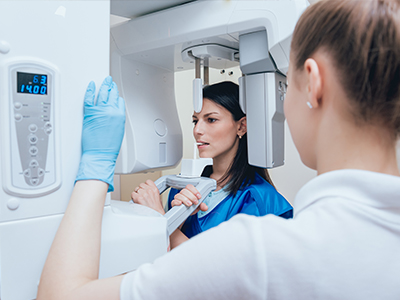
The imaging process itself is brief and generally comfortable. A small sensor or plate is positioned in the mouth for a few seconds while the exposure is taken. Because the exposure time is short and the system is optimized for efficiency, most patients experience minimal discomfort. For those with sensitive gag reflexes or other concerns, clinicians can adapt sensor placement and technique to improve comfort.
After the image is taken, it will appear on the operatory monitor almost immediately. The clinician can then review the image with the patient, pointing out areas of interest and explaining the next steps. This instant feedback helps demystify findings and supports clear communication about preventive measures or necessary treatment.
Regular, appropriately timed radiographs are an important component of comprehensive dental care. Your dental team will recommend imaging only when it provides meaningful information for diagnosis, monitoring, or treatment planning—consistent with best-practice guidelines and individualized to your oral health needs.
Digital radiography offers a combination of improved image quality, faster service, reduced radiation exposure, and enhanced coordination of care. These features support better-informed clinical decisions and clearer patient communication, which together help achieve more predictable outcomes across preventive, restorative, and surgical dentistry.
When digital imaging is paired with other technologies like intraoral cameras, digital impressions, and 3D imaging, practices can deliver highly integrated care that shortens treatment timelines and increases precision. The end result is a more efficient, transparent, and patient-centered experience.
If you’d like to learn more about how digital radiography is used at our practice in Raleigh or how it may benefit your care, please contact us for more information. Our team is happy to explain the process and address any questions you may have.


Digital radiography uses electronic sensors or phosphor plates to capture X-ray exposures and convert them into digital images that appear on a monitor almost immediately. Unlike traditional film, digital systems eliminate chemical development and provide images that can be adjusted for brightness, contrast and magnification. The speed and flexibility of electronic capture change how clinicians review findings and discuss care with patients.
Because the image is produced as a digital file, it can be archived, duplicated and shared without degrading quality in the way that physical film can. Digital capture also permits immediate comparison with prior studies and quicker collaboration with specialists. These practical differences have made digital radiography a foundational tool in modern dental practice.
Higher-resolution sensors and image-enhancement tools help clinicians detect small cavities, root fractures, bone-level changes and other issues that may be difficult to see during a clinical exam. Adjusting contrast and magnification can reveal subtle findings so problems are identified earlier and more reliably. This improved visibility supports more accurate diagnoses and targeted treatment recommendations.
Digital images also allow for precise measurements that inform restorative and surgical planning, such as crown margins, implant positioning and endodontic anatomy. When combined with other digital tools like intraoral scans and CBCT, radiographs contribute to integrated treatment plans that reduce surprises and increase predictability. Overall, clinicians can plan more conservative and effective care with better information at hand.
Digital radiography typically requires lower radiation doses than conventional film techniques to produce diagnostically useful images, thanks to more efficient sensors and image processing. Dentists and radiographers follow the ALARA principle—keeping exposure As Low As Reasonably Achievable—by using shields, collimation and the minimum number of images needed for diagnosis. These safety practices help ensure that imaging is used responsibly across patient populations.
Certain groups, such as children and pregnant patients, receive individualized imaging plans based on clinical need and established guidelines. Clinicians will balance diagnostic benefit against exposure and choose alternatives or defer imaging when appropriate. If you have specific concerns about radiation, your dental team can explain the precautions that will be taken during your appointment.
The imaging visit is usually brief and straightforward: a small sensor or phosphor plate is placed in the mouth and an exposure is taken for a few seconds while the patient remains still. Because exposure times are short and sensors are thin and flexible, most patients experience minimal discomfort, and clinicians can adapt technique for sensitive gag reflexes or other concerns. The entire process is designed to be efficient and comfortable.
Once the image is captured, it appears on the operatory monitor almost immediately, allowing the clinician to review and explain findings during the same visit. This instant review helps patients understand conditions visually and discuss next steps with their provider. Images are then stored in the patient’s digital chart for future comparison and continuity of care.
Digital radiographs are saved as electronic files within the practice’s secure record system, where they can be indexed and retrieved quickly for comparison over time. Electronic storage eliminates the need for physical film and supports organized archiving, which helps clinicians monitor healing, disease progression and treatment outcomes. File formats and retention follow clinical and regulatory best practices for medical records.
When coordination with a specialist is required, images can be transmitted using secure, encrypted methods that protect patient information during transfer. This secure sharing reduces the need to duplicate exposures and shortens treatment timelines by allowing specialists to review high-quality images before consultations. Access to images is controlled so only authorized providers and staff can view protected health information.
Digital radiography is often used alongside intraoral scanners, digital impressions and cone-beam computed tomography (CBCT) to create a comprehensive digital record of a patient’s oral anatomy. Cross-referencing 2D radiographs with 3D CBCT or digital impression data enhances diagnostic insight and enables precise planning for implants, orthodontics and complex restorative cases. Integration of these technologies supports a more coordinated and predictable workflow.
Combined digital tools also streamline communication with dental laboratories and specialists, allowing clinicians to share accurate data for prosthetic design, surgical guides and appliance fabrication. This interoperability reduces manual steps, shortens treatment timelines and improves the overall consistency of care. Patients benefit from more efficient visits and treatments informed by a complete digital data set.
Yes. Digital radiographs reveal internal and subgingival conditions that cannot be seen during a routine visual exam, including interproximal cavities, bone loss, periapical infections and the internal anatomy of roots. These internal views are essential for diagnosing issues that are otherwise hidden behind enamel or soft tissue. Early detection of such conditions often leads to less invasive treatment and better long-term outcomes.
Regular imaging also allows clinicians to monitor areas of concern over time and to detect changes that warrant intervention. By comparing current and prior images side-by-side, providers can identify progression or healing and adjust care plans accordingly. This longitudinal perspective is a key advantage of digital archives.
The frequency of dental radiographs is individualized and based on each patient’s oral health, risk factors and clinical findings rather than a one-size-fits-all schedule. Patients with active disease, a history of cavities, or ongoing restorative or endodontic treatment may need images more often, while low-risk patients without new symptoms typically require fewer exposures. Clinicians follow professional guidelines to determine the appropriate interval.
Your dental team will recommend imaging only when it is likely to provide information that will influence diagnosis, monitoring or treatment planning. This risk-based approach helps limit unnecessary exposure while ensuring clinically relevant data are available when needed. Digital records make it simple to track imaging history and time future studies appropriately.
Digital imaging systems used in dental practices are integrated with electronic health records and adhere to data security standards that protect patient information. Measures commonly include user authentication, role-based access controls and encrypted storage to reduce the risk of unauthorized access. Regular software updates and secure backup procedures further support data integrity and availability.
When images are shared with outside providers, transmissions are carried out using secure, encrypted channels and only after appropriate patient authorization when required. Practices maintain retention and disposal policies consistent with legal and regulatory requirements to safeguard privacy throughout the lifecycle of the record. Patients who have questions about how their images are handled can request more information from their dental team.
The office of Dr. Aaron Tropmann & Dr. Gary Oyster uses digital radiography because it provides clearer images, faster results and more efficient coordination of care than traditional film-based methods. These advantages support earlier detection of dental problems, more precise treatment planning and streamlined visits for patients. The combination of image enhancement and immediate review helps clinicians and patients make informed decisions together.
In practice, digital radiography reduces processing time and chemical waste, improves record-keeping and facilitates secure sharing with specialists when needed, all of which enhance the patient experience in Raleigh and the surrounding communities. If you would like to learn more about how digital imaging fits into your care plan, your dental team can explain the process and answer specific questions during your visit.

Ready to book your next appointment or have a question for our team? We're here to help.
Connecting with our team is simple! Our friendly staff is here to help with appointment scheduling, answer any questions about your treatment options, and address any concerns you may have. Whether you prefer to give us a call, send an email, or fill out our convenient online contact form, we’re ready to assist you. Take the first step toward a healthier, brighter smile – reach out to us today and experience the difference compassionate, personalized dental care can make.

We serve the Raleigh-Durham area, including Cary, Apex, Chapel Hill, Morrisville, Wake Forest, North Hills, North Raleigh and more.
Back to top