
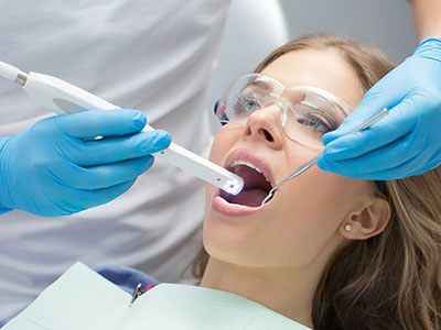
An intraoral camera is a compact, pen-sized imaging device that captures clear, full-color photos and video of the mouth in real time. Unlike a standard camera, it is designed specifically for the oral environment, providing close-up views of teeth, gums, and other soft tissues that are difficult to see with the naked eye. The images appear instantly on a monitor, giving both clinician and patient a shared visual reference during an exam.
Because of its small size and angled tip, an intraoral camera can access tight spaces and subtle angles, revealing early signs of wear, cracks, decay, or gum concerns before they become obvious. The high resolution and adjustable lighting built into modern units make these findings easy to document and review. For patients, that means a more transparent and informed visit—what used to be abstract explanations become visible and understandable.
At the office of Dr. Aaron Tropmann & Dr. Gary Oyster, we view the intraoral camera as part of our commitment to clear communication and precise diagnosis. It’s not a replacement for clinical skill, but a powerful tool that enhances examination, record-keeping, and the overall quality of care.
Using an intraoral camera is quick and comfortable. The hygienist or dentist gently guides the slim wand through the mouth while the camera streams images to a chairside monitor. Patients can watch the screen, giving them a real-time look at the areas being discussed. The process is non-invasive, causes no discomfort, and typically adds only a few minutes to a routine exam.
The camera’s built-in LED lighting eliminates shadows and highlights fine detail, so clinicians can spot hairline fractures, enamel defects, or inflamed tissue that might be missed during a cursory visual check. Many systems also offer still-frame capture and short video clips, enabling a comprehensive record of both static and dynamic conditions—for example, how a restoration responds under bite pressure or how a tissue reacts during movement.
After images are taken, your dental team will explain what is visible on-screen and how it relates to your oral health. This immediate visual feedback often helps patients understand recommended next steps and feel more confident about their treatment plan. If additional imaging is needed, the intraoral camera images can be combined with X-rays or 3D scans for a fuller clinical picture.
Intraoral cameras enhance diagnostic accuracy by revealing details that are hard to observe with standard lighting and mirrors. Early caries, cracked tooth syndrome, and worn margins around fillings or crowns become easier to detect and document. This early detection enables clinicians to recommend minimally invasive solutions before problems progress and require more extensive intervention.
For treatment planning, clear images serve as a reliable baseline. Captured photos can be annotated and stored in the patient record, supporting evidence-based decision-making across multiple visits. When a restorative or cosmetic procedure is under consideration, intraoral photos help the dental team plan margins, shade selection, and the sequence of steps with greater precision.
Documentation from intraoral cameras also supports continuity of care. If a patient requires referral to a specialist—such as an endodontist, periodontist, or oral surgeon—shared images accelerate consultation and help ensure the receiving clinician has a detailed visual summary of the concern prior to initial evaluation.
One of the greatest strengths of intraoral imaging is its ability to make dental health tangible for patients. Viewing a clear photo of a problematic area shifts the conversation from abstract descriptions to concrete evidence, which improves understanding and supports informed consent. Patients who see what their clinician sees are better able to participate in decisions about their care.
Visual documentation is also an effective educational tool. Clinicians can point out anatomy, demonstrate how plaque accumulates in certain locations, or show how existing restorations are performing. This helps patients adopt targeted hygiene habits and track improvements over time. Rather than relying solely on verbal instructions, the team can use images to set measurable goals and revisit progress at follow-up visits.
Because the images become part of the medical record, patients can reference them later or share them with other providers as needed. That continuity fosters trust and reduces uncertainty, especially when multiple appointments or staged treatments are involved.
Modern intraoral cameras are designed to integrate seamlessly with digital practice management systems. Photos and videos are saved directly into the patient’s file, tagged with dates and notes for easy retrieval. This digital workflow reduces paperwork, keeps records organized, and supports clear communication between team members during treatment planning and follow-up care.
When collaboration is required, intraoral images can be exported and shared with specialists or dental laboratories to convey exact clinical findings. High-quality visuals improve the accuracy of lab-fabricated restorations and streamline consultations, since the receiving professionals can preview the clinical situation before the patient arrives. This coordinated approach often shortens turnaround times and improves the predictability of outcomes.
Beyond individual cases, aggregated imaging can support preventive programs and quality assurance within the practice. Tracking visual trends across visits helps clinicians identify changes that may warrant earlier intervention or adjustments in oral hygiene strategies, ultimately supporting long-term oral health for patients.
An intraoral camera transforms routine exams into collaborative, evidence-based conversations by making subtle oral health issues visible and easy to document. From improving diagnosis and treatment planning to enhancing patient education and facilitating specialist collaboration, this technology supports more transparent, efficient, and patient-centered care.
If you would like to learn more about how intraoral imaging is used during exams or how it might benefit your treatment, please contact the office of Dr. Aaron Tropmann & Dr. Gary Oyster for more information. Our team is happy to explain the process and answer any questions you may have.


An intraoral camera is a small, pen-sized imaging device designed to capture high-resolution color photos and short videos of the teeth, gums and other soft tissues inside the mouth. The wand contains a tiny camera and LED lighting that illuminate and photograph hard-to-see areas, producing clear images that appear on a chairside monitor in real time. Because the camera is angled and compact, clinicians can access tight spaces and document subtle issues that might be missed during a visual exam.
The device connects to the practice’s computer system so still images and clips can be saved directly into the patient record for reference and comparison. Many systems allow clinicians to annotate images, capture sequences under bite pressure and combine photos with X-rays or 3D scans for a fuller diagnostic view. This integration supports consistent documentation across visits and helps track changes over time.
Using an intraoral camera is quick, noninvasive and generally comfortable for patients. A hygienist or dentist will gently guide the slim wand through your mouth while the camera streams images to a monitor so you can see what the clinician sees. The process typically adds only a few minutes to a routine exam and causes no physical discomfort.
At the office of Dr. Aaron Tropmann & Dr. Gary Oyster the team will explain the images as they appear, pointing out areas of concern and answering questions to help you understand your oral health. Captured photos can be printed or stored in your digital record so you can review them later or share with another provider if needed. This immediate visual feedback often makes treatment options and next steps clearer to patients.
Intraoral cameras enhance diagnostic accuracy by making fine details visible that are difficult to see with the naked eye or a mirror. High-resolution photos reveal early caries, hairline cracks, worn margins around restorations and subtle tissue changes, allowing clinicians to identify problems at an earlier stage. Early detection often enables more conservative, less invasive treatments before conditions progress.
Because images are recorded and time-stamped, clinicians can compare current photos with prior visits to detect gradual changes or the effectiveness of an intervention. This objective visual record supports evidence-based decisions and reduces the chance of missing developing issues. Clear documentation also helps prioritize care based on clinical need rather than guesswork.
High-quality intraoral photos serve as a reliable baseline for planning restorative, cosmetic and periodontal treatments. Clinicians use images to evaluate margins, select shades, determine preparation design and coordinate sequencing of procedures with greater precision. Photos can be annotated to show proposed treatment areas, which helps the dental team and patient agree on goals before work begins.
When laboratory-fabricated restorations are required, images shared with the dental lab convey important clinical details that improve fit, color matching and overall predictability. Storing images in the patient record also makes it easier to revisit planning decisions during multi-visit or staged treatments. The visual record reduces uncertainty and supports consistent clinical outcomes.
Yes. Intraoral cameras use standard optical sensors and LED lighting and do not expose patients to radiation, making them safe for routine use across most patient populations. The procedure is noninvasive and typically causes no discomfort; the wand is the same size or smaller than many common dental instruments. Clinicians take care to work gently and to minimize any gag reflex or sensitivity during imaging.
Certain patients with extreme restricted opening or severe oral soreness may require modified techniques, but in most cases the camera can be used effectively with small adjustments. If an area cannot be imaged directly, the dental team will rely on complementary diagnostic tools such as radiographs or 3D scans. Safety protocols and infection-control measures are followed for every imaging session.
Seeing a clear photograph of a dental issue turns abstract explanations into concrete evidence, which helps patients understand their condition and the rationale for recommended care. Clinicians can point to a specific image to demonstrate plaque accumulation, a failing margin or the extent of tooth wear, making it easier to explain treatment benefits and alternatives. This visual approach fosters more informed discussions and better patient engagement in decisions about care.
Images saved in the patient record also allow patients to track improvements over time and revisit conversations in follow-up visits. When patients can visually confirm progress, they are often more motivated to adopt recommended hygiene or maintenance routines. Visual documentation therefore strengthens informed consent by ensuring patients know what the clinician observed and why a plan is recommended.
Modern intraoral camera systems are designed to integrate directly with digital practice management and imaging software, so photos and videos are saved in the patient’s chart with date and notes for easy retrieval. This seamless workflow reduces paperwork and ensures that visual documentation is linked to clinical findings, restorative plans and follow-up instructions. Organized imaging in the chart supports continuity of care across multiple visits.
When a referral is needed, high-quality images can be exported and shared securely with specialists or dental laboratories to provide an accurate visual summary before the consultation. This pre-visit information helps specialists triage patients, prepare treatment recommendations and shorten appointment times. The office of Dr. Aaron Tropmann & Dr. Gary Oyster leverages this capability to coordinate care efficiently and maintain consistent communication with outside providers.
No. Intraoral cameras complement radiographs and 3D imaging rather than replace them, because each modality provides different clinical information. Cameras excel at documenting surface detail, soft-tissue appearance and restoration margins in color, while X-rays reveal internal tooth structure, bone levels and interproximal decay that are not visible on the surface. 3D scans and cone-beam CT images provide volumetric information necessary for implant planning and complex surgical cases.
Clinicians use a combination of visual images, radiographs and, when appropriate, 3D scans to build a comprehensive diagnostic picture. The intraoral camera adds valuable surface documentation that can be correlated with radiographic findings to confirm the extent of disease and guide treatment. Selecting the appropriate set of tools depends on the specific clinical question and patient needs.
Infection control is a priority when using any intraoral device, and practices follow manufacturer instructions along with standard clinical protocols. Common measures include using disposable barrier sleeves over the wand, disinfecting external surfaces between patients with approved agents, and following sterilization procedures for any removable components that contact oral tissues. Staff training ensures consistent adherence to these steps with every use.
Routine maintenance and software updates keep image quality reliable and the device functioning properly over time. The dental team documents maintenance activities and follows manufacturer-recommended inspection schedules to identify wear or optical degradation early. Proper care protects patient safety and preserves diagnostic accuracy.
Yes, intraoral cameras are well suited for children and anxious patients because imaging is quick, noninvasive and can be used to build familiarity with the dental environment. Allowing a child or anxious adult to view images on a monitor can reduce fear by demystifying what the clinician is observing and creating a collaborative experience. Clinicians can adopt a gentle, stepwise approach to imaging to increase patient comfort and cooperation.
For patients with severe anxiety or limited tolerance, the team may use distraction techniques, shorter imaging sessions or schedule imaging as part of a calm acclimation visit. When needed, intraoral photos can be taken in stages and reviewed gradually so the patient remains comfortable. The result is accurate documentation without adding stress to the appointment.

Ready to book your next appointment or have a question for our team? We're here to help.
Connecting with our team is simple! Our friendly staff is here to help with appointment scheduling, answer any questions about your treatment options, and address any concerns you may have. Whether you prefer to give us a call, send an email, or fill out our convenient online contact form, we’re ready to assist you. Take the first step toward a healthier, brighter smile – reach out to us today and experience the difference compassionate, personalized dental care can make.

We serve the Raleigh-Durham area, including Cary, Apex, Chapel Hill, Morrisville, Wake Forest, North Hills, North Raleigh and more.
Back to top