
Oral cancer is a serious health concern that often goes unnoticed until it reaches an advanced stage. Early detection dramatically improves treatment options and outcomes, which is why routine screening is a critical part of preventive dental care. During a screening, dental professionals look for subtle signs that might otherwise be dismissed, turning a single appointment into an opportunity to catch disease before it progresses.
Screenings are not just for people with symptoms — they are an important safeguard for every adult who visits the dentist. Because many early lesions cause little or no pain, relying on symptoms alone can delay diagnosis. A structured screening performed during regular dental exams helps identify suspicious findings at a stage when intervention is most effective.
Beyond individual benefit, routine oral cancer screening supports broader public health goals by increasing awareness and encouraging patients to address modifiable risk factors. By making screening a standard part of dental care, clinicians reduce the chance that an otherwise treatable condition becomes life-threatening.
Oral cancer can develop anywhere in the mouth and surrounding tissues, but some areas are more frequently affected. The tongue (especially the lateral borders), the floor of the mouth, the tonsils and oropharynx, the gums, and the inner lining of the cheeks are common sites. Lesions on the lips and hard palate can also occur, particularly where sun exposure or other irritants are present.
Certain factors increase the likelihood of developing oral cancer. Traditional risks include tobacco use and heavy alcohol consumption, which together multiply the danger. Men over 50 historically have shown higher incidence rates, though the profile of people affected is shifting due to other causes. Poor nutrition, a history of head and neck radiation, and long-term irritation or chemical exposures may also contribute.
Human papillomavirus (HPV), especially types associated with oropharyngeal cancer, has become a notable risk factor in recent decades. HPV-related cancers often present differently than tobacco- or alcohol-related disease and may affect a younger, otherwise healthy population. Understanding these evolving risk patterns helps dental teams and patients prioritize regular screening.
An oral cancer screening begins with a focused review of your medical and dental history to identify any risk factors or recent changes in health. The clinician will ask about symptoms such as persistent sores, lumps, swallowing difficulty, or changes in sensation. This background helps tailor the examination to each patient’s needs and establishes a baseline for comparison at future visits.
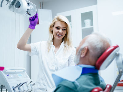
The physical exam includes a careful visual and tactile inspection of the mouth, lips, tongue, gums, and the head and neck region. The dentist or hygienist will look for discolorations, patches, lumps, ulcers, and any asymmetry. Manual palpation of the neck and intraoral tissues can detect enlarged lymph nodes or masses that are not visible on the surface.
Adjunctive technologies can enhance a conventional exam. Tools such as intraoral cameras and digital imaging document findings so clinicians can track changes over time and make informed decisions about follow-up.
When an area of concern is identified, the next steps are thoughtful and evidence-based: observation with short-term recheck, targeted imaging, or referral for biopsy and specialist evaluation. Documentation and clear communication with the patient ensure that suspected lesions are not overlooked and that appropriate care is coordinated promptly.
Knowing which symptoms merit attention helps patients partner effectively with their dental team. Warning signs include a sore or ulcer that does not heal within two weeks, a red or white patch on the oral tissues, or a persistent lump or thickening in the cheek. Changes in the color or texture of the mucosa, unexplained bleeding, or a feeling of numbness or tingling should also prompt evaluation.
Other symptoms outside the immediate mouth can be associated with oral cancers, such as persistent hoarseness, difficulty swallowing, ear pain without an obvious ear problem, or a tightened feeling in the jaw. Any new, unexplained oral symptom that persists should be assessed rather than ignored, especially in people with known risk factors.
Early detection relies on both professional screening and patient vigilance. If you notice any of these signs between dental visits, make an appointment for an evaluation. Timely assessment increases the likelihood that benign conditions are managed conservatively and that malignant conditions are caught at a treatable stage.
If a screening reveals a suspicious lesion, the pathway toward a definitive diagnosis is methodical. Your dentist may recommend monitoring for a short period, refer you for imaging, or arrange a biopsy performed by an oral surgeon or ENT specialist. A tissue biopsy remains the gold standard for diagnosis and guides the next steps in care.
When cancer is confirmed, successful management often involves a team-based approach that may include surgeons, medical oncologists, radiation oncologists, and supportive care providers such as speech and swallowing therapists. Early-stage disease typically allows for less invasive treatment and better functional outcomes, which is why prompt follow-up is so important.
Prevention and risk reduction are central to ongoing care. Practical steps include quitting tobacco, reducing alcohol intake, protecting the lips from sun exposure, and maintaining a balanced diet rich in fruits and vegetables. HPV vaccination is an effective preventive measure against the strains most commonly linked to oropharyngeal cancers and is part of larger public health efforts.
At the office of Dr. Aaron Tropmann & Dr. Gary Oyster, patients can expect careful documentation, clear explanations of findings, and coordinated referrals when needed. Our clinicians emphasize patient education so individuals understand both their screening results and the reasons behind recommended next steps, helping them make informed choices about their health.
In summary, oral cancer screening is a low-risk, high-impact component of routine dental care that helps detect disease early, guide timely intervention, and support long-term oral health. If you have questions about the screening process, risk factors, or what to expect during an exam, please contact us for more information.


Oral cancer screening is a systematic exam of the mouth, lips, tongue, gums and surrounding tissues performed to detect early signs of abnormal or precancerous changes. The goal is to find suspicious lesions or symptoms before they progress, when treatment is typically simpler and outcomes are better. Because many early abnormalities cause little or no pain, screening is a vital element of preventive dental care.
Screening usually occurs as part of a routine dental visit and combines a focused health history with a visual and tactile inspection of the oral and neck regions. Clinicians document findings and compare them with previous exams to identify changes over time. When necessary, further testing or specialist referral is arranged to reach a definitive diagnosis.
All adults should receive an oral cancer screening during their regular dental exams, as early lesions can occur in people without symptoms or obvious risk factors. Patients with known risk factors — such as tobacco use, heavy alcohol consumption, prior head and neck radiation, or a history of oral lesions — may be monitored more closely. Screening frequency is individualized and may be increased when clinicians identify suspicious findings or elevated risk.
Dental teams establish a baseline exam for new patients and track any changes at subsequent visits, which helps guide follow-up intervals. Younger adults with risk factors such as HPV exposure should also be screened routinely. Open communication about medical history and new symptoms helps clinicians tailor screening schedules to each patient’s needs.
Traditional risk factors include tobacco use in any form and heavy alcohol consumption, which together markedly increase the likelihood of oral cancer. Prolonged sun exposure to the lips, poor nutrition, a history of head and neck radiation, and chronic irritation from ill-fitting dental appliances or chemical exposures also elevate risk. Occupational exposures and certain immunosuppressive conditions may further contribute.
Human papillomavirus (HPV), particularly strains linked to oropharyngeal cancer, has become an increasingly important risk factor and may affect younger, otherwise healthy individuals. Understanding a patient’s full risk profile helps clinicians focus screening efforts and recommend preventive steps such as tobacco cessation and HPV vaccination where appropriate.
A modern screening begins with a focused review of medical and dental history along with questions about symptoms such as persistent sores, lumps, swallowing difficulty, or changes in sensation. The clinician performs a careful visual inspection and manual palpation of the lips, oral mucosa, tongue, floor of mouth, gums, and the lymph nodes in the neck. Detailed documentation and intraoral photography are often used so clinicians can compare findings over time.
Adjunctive tools can supplement the clinical exam; examples include tissue fluorescence devices such as intraoral cameras and targeted imaging to clarify suspicious areas. These technologies enhance visualization and record-keeping but do not replace clinical judgment. When an area appears concerning, clinicians discuss next steps which may include short-term monitoring, imaging, or referral for biopsy.
You should seek prompt evaluation for any sore or ulcer in the mouth that does not heal within two weeks, persistent red or white patches, or a lump or thickening in the cheek or under the tongue. Other warning signs include unexplained bleeding, sudden numbness, or changes in the color or texture of oral tissues. Because these signs can indicate a range of conditions, timely assessment helps distinguish benign issues from those requiring further investigation.
Symptoms outside the mouth such as persistent hoarseness, difficulty swallowing, unexplained ear pain, or a growing neck mass can also be related to cancers of the oral cavity and oropharynx. If you notice new, unexplained symptoms between dental visits, contact your dental provider for an evaluation. Early assessment increases the likelihood of detecting treatable conditions and avoiding delayed diagnosis.
If a clinician identifies a suspicious area, the first steps are careful documentation and a clear discussion of possible causes and next actions. Options commonly include short-term observation with a recheck, targeted imaging to define the lesion, or referral to an oral surgeon or ENT specialist for further evaluation. A tissue biopsy is often recommended when the appearance or behavior of a lesion raises concern.
A tissue biopsy remains the gold standard for diagnosis and helps guide treatment planning. If cancer is confirmed, management typically involves a multidisciplinary team that may include surgeons, medical oncologists, and radiation specialists. Your dental team can help coordinate referrals, communicate findings to specialists, and support you through each step of the diagnostic and treatment process.
Adjunctive tools such as brush biopsy kits and enhanced imaging can improve the clinician’s ability to detect abnormalities and document lesions. These technologies make some subtle changes easier to see and help guide decisions about which areas warrant closer attention or biopsy. However, they are screening aids rather than definitive diagnostic tests.
Because adjunctive tests can produce false positives and false negatives, clinicians interpret results in the context of the full clinical exam and patient history. Suspicious findings identified with adjunctive methods typically require histologic confirmation by biopsy. The most effective approach blends experienced clinical examination with selective use of adjunctive tools to optimize early detection.
While not all cases can be prevented, many oral cancers are associated with modifiable risk factors that patients can address to reduce risk. Key prevention strategies include quitting tobacco, limiting alcohol consumption, protecting the lips from excessive sun exposure, and maintaining a balanced diet rich in fruits and vegetables. HPV vaccination is a highly effective preventive measure against the strains most commonly linked to oropharyngeal cancers.
Regular dental visits with routine screening and patient education are central to prevention efforts because they enable early detection and support behavior change. Clinicians can provide resources and referrals for tobacco cessation, vaccination guidance, and nutritional counseling. Combining risk reduction with vigilant screening offers the best protection against late-stage disease.
At our Raleigh practice patients receive clear explanations of findings, documented records and a written plan for next steps when a suspicious lesion is found. We prioritize timely communication and, when appropriate, arrange targeted imaging or prompt referrals to oral surgery, ENT or oncology specialists so that diagnostic steps are not delayed. Our team also explains what to expect during each stage and provides educational resources to help patients make informed decisions.
When biopsy or specialist care is required, we coordinate information transfer and follow the patient’s progress to ensure continuity. Supportive care needs, such as nutrition counseling or speech and swallowing therapy, are discussed as part of comprehensive planning. The goal is to guide patients through the diagnostic process with compassion, clear information and efficient coordination of care.
Between visits, perform simple self-checks of your mouth by looking for persistent sores, lumps, patches of discolored tissue or any new or unusual changes. Report any suspicious findings to your dental provider promptly rather than waiting for the next scheduled exam. Maintaining good oral hygiene and promptly addressing chronic irritation from broken teeth or ill-fitting appliances also reduces the chance that problems are overlooked.
Adopting healthy habits such as avoiding tobacco, moderating alcohol intake, protecting your lips from sun exposure and staying current with vaccinations supports long-term oral health. Keep your dental team informed about changes in your medical history, medications or new symptoms so that screening can be personalized and effective. Early action and open communication are powerful tools for detecting disease at a treatable stage.

Ready to book your next appointment or have a question for our team? We're here to help.
Connecting with our team is simple! Our friendly staff is here to help with appointment scheduling, answer any questions about your treatment options, and address any concerns you may have. Whether you prefer to give us a call, send an email, or fill out our convenient online contact form, we’re ready to assist you. Take the first step toward a healthier, brighter smile – reach out to us today and experience the difference compassionate, personalized dental care can make.

We serve the Raleigh-Durham area, including Cary, Apex, Chapel Hill, Morrisville, Wake Forest, North Hills, North Raleigh and more.
Back to top