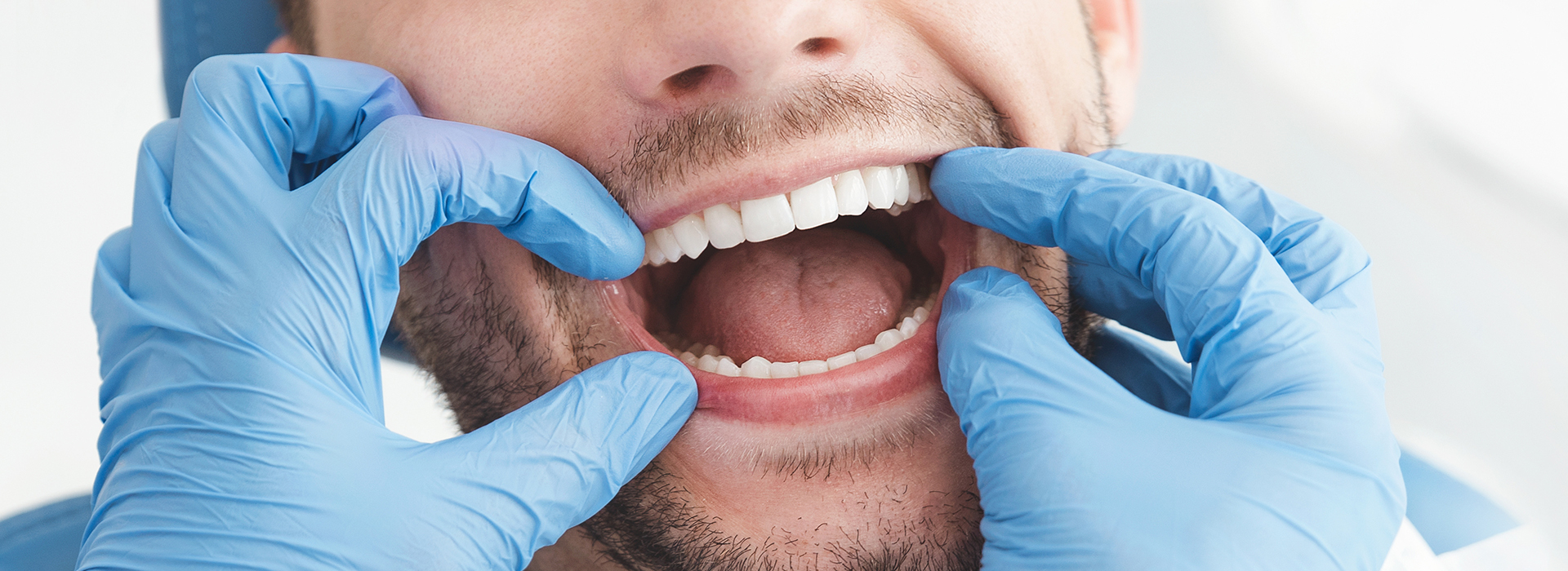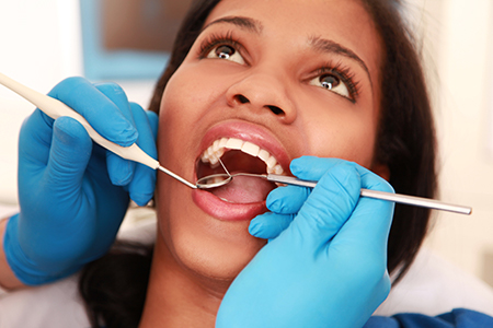
At the office of Dr. Aaron Tropmann & Dr. Gary Oyster, we prioritize prevention and clear communication as the cornerstones of lasting oral health. A well-conducted oral exam is more than a checklist — it’s an opportunity to understand how your mouth is functioning today and what steps will keep it healthy tomorrow. During each visit, our team combines careful observation, modern diagnostics, and patient education so you leave with a clear sense of your needs and next steps.
Your first oral exam with us creates a detailed baseline that guides all future care. We start by reviewing your medical history and any medications or lifestyle factors that influence oral health, then discuss your concerns, goals, and any symptoms you may be noticing. This conversation helps us tailor the exam to your individual situation and ensures we don’t miss anything relevant to your overall care.
Next comes a focused clinical evaluation of the teeth, gums, tongue, and surrounding soft tissues. We look for signs of decay, early gum disease, wear patterns from grinding or clenching, and issues with jaw alignment or bite. Where appropriate, we assess temporomandibular joint (TMJ) function and screen the head and neck for any irregularities that could warrant further investigation.
Diagnostic imaging and other tests are used selectively to expand what we can see by eye. If x-rays or three-dimensional imaging are recommended, we explain why they add value and how the information will influence treatment planning. At the end of the initial assessment, you’ll receive a clear explanation of findings and the most important next steps — whether that’s simple maintenance, targeted preventive care, or a staged treatment plan.

An oral exam often reveals clues about conditions that extend beyond the mouth. Changes in gum tissue, persistent ulcers, dry mouth, or unusual lesions can reflect systemic issues such as nutritional deficiencies, autoimmune disorders, or side effects of medications. By examining the oral cavity in context, we can help identify signs that may warrant coordination with a medical provider.
Research continues to show connections between oral inflammation and broader health concerns. While an exam can’t diagnose every systemic disease, it can flag warning signs that lead to earlier detection and intervention. Our approach is to interpret oral findings thoughtfully and offer appropriate recommendations for follow-up when we suspect a link to overall health.
Part of our role is educating patients about these connections so they can make informed choices. Simple changes in hygiene, diet, or medication management — discussed during an exam — can sometimes reduce risk or improve symptoms. When necessary, we document findings and communicate clearly with other healthcare professionals to support coordinated care for the whole person.
Routine exams and professional cleanings are the most effective way to prevent small problems from becoming large ones. Even diligent brushing and flossing at home can miss deposits that harden into tartar; during a professional cleaning, our hygienist removes these deposits and polishes the teeth, reducing the bacteria that cause decay and gum disease.
Regular visits give us the chance to monitor changes over time. Subtle shifts in tooth position, the development of staining, or early areas of gum recession are easier to address when they’re caught early. For many patients, twice-yearly visits are sufficient; for others with higher risk factors, more frequent monitoring may be recommended. We base that recommendation on objective findings and your personal risk profile.
Checkups are also an opportunity for practical coaching. Our team demonstrates techniques for brushing and interdental cleaning tailored to your anatomy and any appliances you use, such as braces or removable prosthetics. We discuss diet-related risks and habits that accelerate wear or sensitivity, and explain how preventive treatments can fit into a realistic home care routine.
For children, routine exams play a key role in establishing healthy habits and tracking development. We watch how primary and permanent teeth emerge, screen for early orthodontic concerns, and provide age-appropriate guidance so young patients grow into healthy adult smiles.
Visual inspection is vital, but radiographs reveal the structures beneath the surface. X‑rays and digital imaging let us evaluate root health, bone levels, the presence of developing or impacted teeth, and areas of decay that aren’t visible clinically. When used judiciously, imaging improves diagnostic accuracy and treatment outcomes.
Modern digital radiography reduces exposure while improving image clarity and workflow. Digital images are available immediately, can be enhanced for better interpretation, and are easy to store and share securely when coordination with other providers is needed. We follow accepted guidelines to recommend the right images at the right intervals, balancing diagnostic benefit and safety.
We’ll always explain why a particular image is recommended and how it will be used. Whether assessing a possible infection, evaluating bone for an implant, or monitoring the progression of a lesion, clear imaging helps us plan treatment precisely and communicate options to you in understandable terms.
Limited radiation exposure with faster results
Immediate viewing and enhanced image manipulation
Safe electronic storage and easier coordination of care
Environmentally friendly workflow with no chemical processing
Images become part of your permanent record for long-term comparison

Different imaging techniques serve different diagnostic purposes. Small intraoral films are ideal for evaluating individual teeth and detecting early cavities, while panoramic images provide a broad overview of the jaws and developing dentition. When three-dimensional detail is needed, cone-beam CT (CBCT) gives us precise spatial information for surgical planning and complex restorative work.
Here are some common imaging types and what they reveal:
Periapical view — focused on a single tooth and its root, useful for detecting root infections and assessing supporting bone.
Bitewing — captures the crowns of adjacent teeth and is a sensitive way to find early interproximal decay.
Full mouth series — a combination of intraoral views that provides a comprehensive baseline of dental health.
Panoramic film — a broad 2D image of both jaws, useful for seeing eruptions, impactions, and overall jaw structure.
Cephalometric film — a profile view used primarily in orthodontic assessment and growth evaluation.
CBCT imaging brings three-dimensional clarity when planning implants, evaluating complex anatomy, or assessing pathology that cannot be fully understood in two dimensions. As with all tools, we recommend CBCT only when the additional information will materially improve diagnosis or treatment planning.
By combining a careful clinical exam with the appropriate imaging, we deliver balanced, evidence-based recommendations. Our goal is to give you a straightforward path forward that preserves function, prevents disease, and maintains the appearance you want from your smile.
In summary, a comprehensive oral exam is the foundation of smart, patient-centered dentistry. Regular assessments let us catch problems early, coordinate with your medical team when needed, and provide targeted preventive care. If you have questions about what to expect during an exam or whether additional imaging is right for you, please contact us for more information.

An oral exam begins with a brief review of your medical history and any medications or symptoms you are experiencing. The clinician then performs a systematic visual and tactile evaluation of the teeth, gums, tongue, cheeks, and other soft tissues to check for decay, gum disease, unusual lesions, wear patterns, and signs of occlusal stress. The exam often includes a check of jaw movement and temporomandibular joint function and a palpation of the head and neck to screen for enlarged lymph nodes or other irregularities. If additional information is needed, the team will recommend appropriate imaging or tests and explain why those tools will help with diagnosis.
After the clinical evaluation, the dentist or hygienist will summarize the findings and outline recommended next steps, which may range from routine preventive care to a staged treatment plan. Patient education is a core part of the visit, so you can expect guidance on home care techniques, behavior changes, and ways to manage any symptoms. Staff will also document baseline measurements and images so future visits can track changes over time. The goal is to leave you with a clear understanding of your oral health status and practical steps to maintain or improve it.
Regular oral exams allow clinicians to catch small problems before they become larger and more complex. Early detection of cavities, gum inflammation, and wear-related damage makes treatment simpler and more predictable, and professional cleanings remove deposits that home care may miss. Exams also provide an opportunity to monitor changes in tooth position, restorations, and soft tissues so that trends can be addressed proactively. Over time, this preventive approach helps preserve function and reduces the risk of urgent problems.
Consistent monitoring also creates a reliable record that helps tailor care to your individual risk profile, including how often you should be seen. Based on objective findings such as bone levels, plaque accumulation, and medical risk factors, the team may recommend more frequent visits for some patients and standard recall for others. This personalized cadence supports long-term oral health and efficient use of clinical resources. Education and reinforcement of home-care techniques further increase the benefits of regular visits.
For many adults and children, a routine exam every six months provides an effective balance between prevention and practicality. That interval supports regular professional cleaning, timely detection of new decay, and monitoring for early signs of gum disease. However, the ideal frequency depends on individual risk factors such as a history of periodontal disease, active decay, tobacco use, certain medical conditions, or medications that cause dry mouth. Patients with higher risk profiles may need recall visits every three to four months or on a schedule recommended by the dentist.
When you come for an exam, the dental team will assess your oral health and discuss recall timing based on measurable findings and your personal circumstances. They will document the rationale for the recommended interval so you understand why a particular schedule is suggested. If your health status or habits change, the team can adjust the frequency to reflect new needs. The goal is to choose a cadence that optimizes prevention while fitting your lifestyle.
Yes. A routine oral exam typically includes a visual and tactile screening for suspicious lesions or abnormalities that could suggest oral cancer. The clinician inspects the lips, tongue, floor and roof of the mouth, inner cheeks, and throat for persistent sores, white or red patches, indurated areas, or other unusual findings, and palpates the neck to check for abnormal lymph nodes. Because early detection significantly improves outcomes, any suspicious area is documented, photographed when appropriate, and monitored closely.
If a lesion appears suspicious, the team will explain the next steps, which may include a short-interval recheck, referral to a specialist, or biopsy when clinically indicated. The screening is risk-based and more frequent for patients with known risk factors such as tobacco or heavy alcohol use and certain viral exposures. Throughout the process, clinicians provide clear explanations so patients understand the reason for follow-up and the signs to watch for between visits.
Dental x-rays are recommended when they add diagnostic value beyond visual inspection, such as detecting early interproximal decay, assessing root health, or evaluating bone levels around teeth. The dentist will consider your clinical findings, caries risk, age, and previous imaging when deciding which views are appropriate and how often to take them. Modern digital radiography minimizes radiation exposure and provides high-quality images that can be enhanced for clearer interpretation. You will always be told why a specific image is useful and how it will inform your care.
Three-dimensional imaging such as cone-beam CT is reserved for cases where spatial detail materially affects treatment planning, for example when planning implants, evaluating complex anatomy, or assessing pathology that cannot be fully understood in two dimensions. CBCT is not used routinely and is recommended only when the additional information will change the clinical approach. The practice follows accepted guidelines to balance diagnostic benefit with patient safety and discusses alternatives when appropriate.
The oral cavity often reflects changes related to overall health, so an exam can reveal clues that warrant medical attention. Symptoms such as persistent dry mouth, chronic ulcers, unusual oral pigmentation, swollen or bleeding gums, and delayed healing may point to systemic conditions like diabetes, nutritional deficiencies, autoimmune disorders, or adverse medication effects. Because the exam considers the mouth in context with your medical history and medications, clinicians can identify patterns that suggest a broader health concern.
When a possible systemic link is suspected, the dental team documents findings clearly and communicates recommendations for follow-up with your medical provider. This coordination supports more timely diagnosis and treatment of underlying conditions and helps ensure that dental care is safe and effective within the context of your overall health. Patient education about oral–systemic connections is also emphasized so you can take practical steps to reduce risk and support general well-being.
Before an exam, it is helpful to provide an up-to-date medical history, a list of current medications and supplements, and any recent changes in health or symptoms. Tell the team about dental concerns, pain, sensitivity, or functional issues such as difficulty chewing or jaw pain, and share information about habits like tobacco or alcohol use and sleep-related breathing problems. If you are a new patient, bringing previous dental records or recent radiographs can speed diagnosis and avoid unnecessary repeat imaging.
Also mention any allergies, bleeding disorders, or medical devices such as pacemakers that might affect care, and let staff know if you experience dental anxiety so appropriate accommodations can be made. Clear communication helps the clinician tailor the exam and any recommended follow-up to your needs. Finally, prepare any questions you want addressed so the visit is as informative and efficient as possible.
Children’s oral exams focus on growth and development as well as preventing disease. Clinicians assess how primary and permanent teeth are erupting, look for early signs of cavities or developmental enamel defects, and screen for habits like thumb-sucking or mouth breathing that can affect alignment. Early exams also identify the need for interceptive orthodontic evaluation and help determine the timing of preventive treatments such as fluoride or sealants.
Exams for young patients include age-appropriate education for both the child and caregiver, with practical coaching on brushing, flossing, and diet. The visit is structured to build a positive relationship with dental care and reduce anxiety, while tracking developmental milestones over time. Based on findings and risk, the team will recommend a recall schedule that supports healthy dental development into adolescence.
CBCT is indicated when three-dimensional detail will materially affect diagnosis or the safety and success of treatment, such as in implant planning, locating impacted teeth, evaluating complex root anatomy, or assessing suspected pathology. It provides precise spatial information that two-dimensional images cannot capture, which can be critical for surgical planning and risk assessment. The decision to use CBCT is made judiciously and only after considering whether the additional information will change the proposed clinical approach.
When CBCT is recommended, clinicians explain the reason, what the scan will reveal, and how the information will be applied to your care plan. The practice follows principles to minimize exposure and uses the smallest field of view necessary to answer the clinical question. Patients are given an opportunity to ask questions and to review the images with the clinician so they understand how the scan influences treatment choices.
After the exam you should receive a clear summary of findings and a recommended plan of care that prioritizes the most important next steps. This plan may include preventive measures, a recommended schedule for professional cleanings, targeted treatments such as fillings or periodontal therapy, and any suggested imaging or specialist referrals. The team will explain the rationale for each recommendation and provide instructions for at-home care to support healing and prevention.
At the office of Dr. Aaron Tropmann & Dr. Gary Oyster, documentation from the exam becomes part of your permanent record so progress can be monitored over time. If coordination with a medical provider or dental specialist is helpful, the practice will communicate findings and facilitate referrals as appropriate. Follow-up appointments are scheduled based on clinical need and patient preference to ensure continuity of care and the best possible outcomes.

Ready to book your next appointment or have a question for our team? We're here to help.
Connecting with our team is simple! Our friendly staff is here to help with appointment scheduling, answer any questions about your treatment options, and address any concerns you may have. Whether you prefer to give us a call, send an email, or fill out our convenient online contact form, we’re ready to assist you. Take the first step toward a healthier, brighter smile – reach out to us today and experience the difference compassionate, personalized dental care can make.